Tag Archives: Immunology

Skin Diseases, Disruption and Immune Dysregulation, 2/10/26

TERMS
Epidermis = Outer most layer of the skin, serves as a protective barrier.
Dermis = Middle layer of the skin, actively growing and providing nutrients to the skin.
Sebaceous Glands = Glands at the base of hairs that release an oily substance to the surface to waterproof and protect the outer epidermis layer.
Eczema = Group of skin diseases featuring dry, red, itchy, oozing skin that may be caused by many factors; irritants (chemical or environmental), inflammation (Atopic Dermatitis), glandular disfunction, veinous disorders or neurological dysfunction.
Atopic Dermatitis = Group of inflammatory driven skin diseases, often identified by dry, red, itchy, oozing skin often in inner joint areas. This is a type of Eczema.
Psoriasis = Group of inflammatory skin diseases, often identified by areas of white/pink scaly and flaky plaques of skin often on outer joint surfaces. Can cause systemic inflammation.
Psoriatic Diseases = Term given to a class of more severe Psoriasis that involves other tissue such as joints, eyes and intestines.

Intro to the Skin
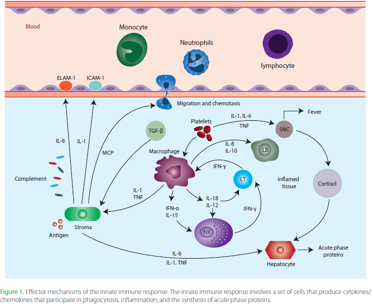
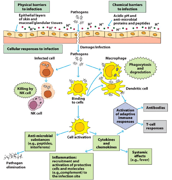
The term “Skin” most often refers to the outer layer of cells that cover and protect most of our body (and other mammals too). Though in reality the skin is a very complex organ that contains not just this outer barrier, but a complex network of cells that contribute to physically shielding us from injury, sensing environmental changes, regulating our body and comprising complex immune functionality that serves to both repair damage and ward off any invader that might break through. Unfortunately, there are times when this complex multi-faceted system becomes dysfunctional or is coopted, inducing disease. In this installment of Making Science Accessible I’ll be discussing the basics of skin anatomy (structure) and function, followed up with deeper dive into two major skin diseases, Atopic Dermatitis (common type of Eczema) and Psoriasis. Discussing how they’re similar, how they differ and why these relationships can alter how one seeks treatment and reprieve from the disease.

Most people see The Skin as a single layer of protection on the outside of our body, but it’s really much more complex than that. The skin is comprised of three major layers; outer Epidermis (that we see), Dermis (middle) and the underlying fatty layer. The Epidermis is what most people are familiar with, serving as the physical barrier to the outside world, protecting us from contact injury, UV damage (melanin) and infection. This outer layer is constantly being rebuilt by dividing keratinocyte (skin) cells that slowly push upwards until they reach the surface as a tightly woven network of dead cells, creating a solid barrier. The second layer, the Dermis, is the thickest part of the skin and is where most of the action happens. The Dermis contains most of the blood vessels that supply the skin with nutrients, lymph vessels that house our immune cells, hair follicles, nerve endings and sweat/oil glands. The innermost layer of the skin is the Subcutis, made up of a network of collagen and fat cells that serve to insulate and absorb shock, protecting the underlying tissue and organs. Dysfunctions can occur within any layer of the skin (cancer, structural, inflammatory), though this writing will primarily focus on immune related diseases that originate in the Dermis, subsequently affecting the surrounding tissue.

Eczema vs Psoriasis
The two most common forms of inflammatory skin disease are Eczema and Psoriasis. On the surface (drum roll) they would appear to be very similar, but once you dig deeper into their contributing disease mechanisms, there are noticeable differences. First, I’ll do a brief compare and contrast of the two, before diving into a more detailed discussion about each individual disease class, it’s contributing mechanisms, clinical signs and treatment options. Eczema is a broader term that is medically used to describe a group of different skin disorders that all manifest similarly. Eczema can be caused by underlying immune dysfunction (Atopic Dermatitis), contact with irritants such as chemicals and allergens (ie Poison Oak/Ivy), nerve irritation, defects in the Sebaceous glands (severe cases of dandruff) and vein disorders. While all of these can cause similar dry, red, itchy and oozing skin rashes, I’ll be focusing mostly on Atopic Dermatitis, the most common chronic form of Eczema.

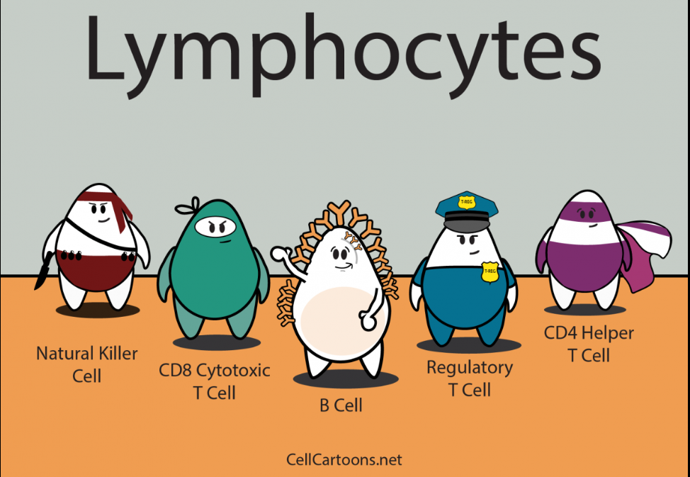
While both Atopic Dermatitis and Psoriasis lead to Epidermal and Dermal irritation and barrier damage, the specifics of their outward appearance are often what’s used for differential diagnosis. The differences in their external appearance are often caused by dysfunction of different immune and cellular mechanisms within the body, but there is also much overlap between the two diseases as well. Both can have genetic mutations that predispose one to developing the disease, and this linkage is typically stronger the more severe disease an individual suffers. Of the genes that are commonly dysregulated in both diseases, approximately 80% overlap, but that also means that 20% of what is dysfunctional is different. Both diseases are commonly driven by hyper-activated Tcells in the Dermis, leading to excessive inflammation, Epidermal barrier breakdown (ie skin damage) and overall dysfunction. But when one drills down at a more functional level, Atopic Dermatitis is primarily driven by Type 2 Tcells (Th2), while Psoriasis is driven by Th17/Th22 Tcells (that produce signaling molecules IL-17 and IL-22). These differences in signaling and function recruit different parts of the immune system to the skin, meaning that the primary cells involved in the inflammation and dysfunction can differ. Though the clinical reality is that both Atopic Dermatitis and Psoriasis exist on a spectrum, where both the clinical manifestations and the underlying cellular mechanisms can be quite variable from individual from individual. Meaning that while general trends in appearance and causes might exist, each case (patient) has its own unique fingerprint. In fact, there is growing evidence that some patients can suffer from both diseases, with them either occurring simultaneously or alternating (known as Psoriatic Dermatitis). In these cases, the immune dysfunction signatures and clinical signs will overlap or alternate, further confounding diagnosis and treatment. In the next few sections I will be discussing each disease in isolation, though you may notice a lot of overlap between the causes and treatments, further showing how complex and intertwined immune related diseases can be in general.

Classical Immune Cell activation pathways in Atopic Dermatitis (AD), Psoriasis (PSO) and overlapping disease. Targeted therapies aim to block pieces of each respective chain.
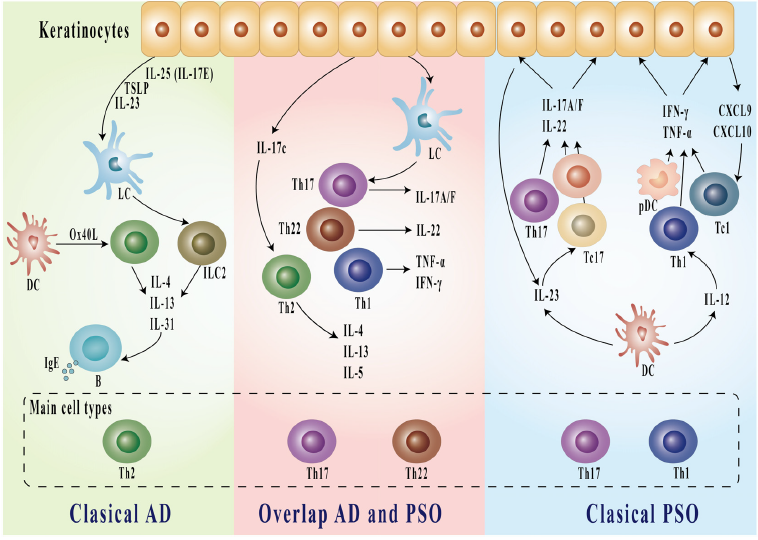
Figure 1, Li et al. Beyond the Dichotomy: Understanding the overlap between Atopic Dermatitis and Psoriasis.

Atopic Dermatitis/Eczema, Disease Biology, Variability and Treatment
While the term Eczema is often used interchangeably to describe Atopic Dermatitis (one form of Eczema), medically it’s a classification for a group of dermatitis skin disorders with similar clinical appearance, but different underlying mechanisms. Skin disorders such as Seborrheic Dermatitis (sebaceous gland dysfunction), Contact Dermatitis (chemical or allergic irritant induced) and other less common diseases all fall under the same Eczema umbrella. Seborrheic Dermatitis is associated with a dysfunction of sebaceous glands, which produce an oily substance that serves to lock in skin moisture and prevent infections. When these glands don’t produce the appropriate amount of fluid the skin can become irritated, dried out and/or prone to infection by fungi (commonly Malassezia spp). This dysregulation leads to inflammation and the dry itchy skin rashes commonly associated with Eczema. For Seborrheic Dermatitis, appearance wise this often manifests as more severe cases of dandruff or facial Eczema. Common treatments include anti-fungal and anti-inflammatory creams and shampoos to treat the condition, which is typically well managed. The Contact Dermatitis form of Eczema is commonly associated with skin rash formation driven by an allergic reaction in response to an external sensitivity, such as a chemical or allergen. Common forms of Contact Dermatitis are Poison Oak/Ivy reactions, reactions to detergents and other household chemicals. Treatment can be as simple as cleaning the affected area and letting the irritation calm down or include topical and oral anti-inflammatory therapies in more severe cases. As one who suffers from both mild Contact Dermatitis to chemical irritants and severe skin reactions to Poison Oak/Ivy I can attest to how painful and debilitating the condition can be, but also how well it’s managed with special cleaning agents and steroids.

While Seborrheic and Contact Dermatitis are typically well controlled with treatment, Atopic Dermatitis (AD) can be a more problematic disease due to variability in the underlying causes and more inconsistency in treatment efficacy. Mutations in the skin structural proteins (filaggrin), Th2 signaling pathways (IL-4 and IL-13) and other immune activators have been linked to increased risk for developing AD. These are all linked to increased activity of the Th2 pathway, which is the primary driver of AD disease and skin damage. Th2 responses are directed by a specific subset of Tcells that are typically activated by allergens and parasites. When properly regulated, Th2 immune responses help to clear these external trouble-makers from our body by activating IgE Antibody production and Mast Cells and Eosinophil cells. These serve to wall off the foreign entities and then break them down to be removed from the skin. In AD these responses become inappropriately activated, and the cycle of overly exuberant Th2 mediated immune activity drives degradation of the epidermal barrier (skin), allowing additional allergens and bacteria (such as Staph aureus and Streptococcus, common in AD patients) to penetrate the skin, creating additional inflammation and damage that can become self-propagating. In some cases the skin damage comes first, in others the underlying immune activity, regardless the end result is a predominantly dysregulated Th2 immune response in the skin that drives continual activation, damage and irritation. While the dysregulated Th2 immune response is the most prominent immune feature in AD, high levels of activity in Th17 (IL-17 signaling Tcells) and Th22 (IL-22 signaling Tcells) cells can be contributors to disease. These Tcells use different signaling pathways to activate microbial defense mechanisms (Th17) or to activate skin proliferation and healing (Th22). Just as with the Th2 immune response, when properly regulated and activated in moderation they help protect and heal our body, but when over activated the Th17 pathway can cause additional tissue damage and the Th22 response can cause skin thickening and scarring, rather than normal healing. In summary, AD is typically caused when normal skin immune cell function becomes dysregulated and over-activated, which damages the Epidermis, causing irritation, which allows more foreign molecules to get under our skin, perpetuating the cycle of dysfunctional immune activation.

Circular cycle of skin damage and inflammation that occurs during Atopic Dermatitis and the associated pathways for treatment.
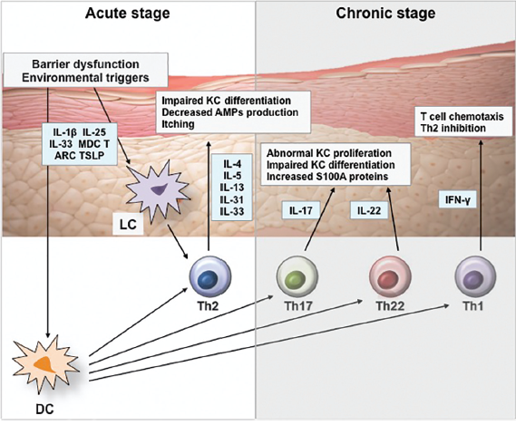
Figure 1, Kim et al. Pathophysiology of Atopic Dermatitis: Clinical Implications.

Because of the varied causes and contributing factors to AD and Eczema, optimal treatment for controlling skin damage and inflammation can vary depending on the individual case. Treatments come in four main categories: generic skin supportive therapies, topical anti-microbials, topical anti-inflammatory treatment and systemic anti-inflammatory treatment. For more mild forms of AD and Eczema basic topical treatments to clean, hydrate and support the skin can help mitigate some of the itching and irritation, though this is more of a supportive than specific disease targeting treatment. These can include lotions/creams to moisturize the skin, UV/sun exposure and regular cleaning of the affected area. Antibiotics and antifungals (mostly topical) can also be helpful to mitigate some of the potential infection risk associated with certain bacteria and fungi, reducing the infection induced immune activation. While these won’t treat the underlying immune dysfunction, they can help reduce severity and immune activation. The next classification of treatments, topical anti-inflammatories, are effective for many cases of AD/Eczema in addition to other skin diseases (like Psoriasis). Broad-spectrum topical anti-inflammatories include common compounds such as corticosteroids, cyclosporin and methotrexate, all of which serve to generally suppress immune responses, with no specificity for cell type or pathway. This broad-spectrum efficacy is both their strength and their weakness. It’s a strength because they can work against any underlying immune dysfunction (where overactivation occurs) regardless of the pathway. So even if one doesn’t know the specific immune dysfunction, these broad-spectrum immunosuppressants can help. The weakness is, because they don’t target only the dysfunctional immune pathways, other protective systems in the body can also be suppressed AND the dysfunctional pathway can even be suppressed too much, leading to susceptibility to other infections. These are common risks and side effects of taking broad-spectrum anti-inflammatories for prolonged durations and is why they aren’t the ideal therapeutics for long term maintenance of a disease, but can be helpful to reduce short term flares or acute disease.

The last category of treatments for AD/Eczema are systemic anti-inflammatory treatments. While this can include broad acting anti-inflammatories like those mentioned above, for AD/Eczema systemic treatments usually leverage cell type or pathway specific therapies. Things such as monoclonal antibodies and drugs that are specifically designed to bind to one cell, receptor or signaling molecule. These types of targeted therapeutics can be great because treatment can target only the dysfunctional parts of the immune system without reducing the properly functioning areas. Treatments exist to target IL-4 and IL-13 (Th2 pathway), IL-17 and IL-23 (Th17 pathway), IL-12/23 (Tcell activation) and JAK/STAT (generic immune activation), all of which have been shown to help people suffering from AD/Eczema depending on the immune mechanisms that are dysfunctional. The challenge with these therapeutics is the variability of AD/Eczema disease in each person and the challenge of identifying the exact mechanisms of dysfunction for a single individual. Each targeted treatment will help some individuals more than others, and while there is testing that can help identify these pathways, it’s not highly utilized and readily available to most patients. I’ll discuss this more in the next post about “Personalized Medicine and Immune Signatures”.

Psoriasis and Psoriatic Arthritis, Disease Biology, Variability and Treatment
Like Eczema (and Atopic Dermatitis), Psoriasis is a skin disease characterized by Epidermal damage, irritation and underlying immune dysregulation, but with some striking differences in both its external and internal manifestation. The cellular hallmark of Psoriasis is keratinocyte dysfunction (these are the cells that make up our skin), primarily caused by the underlying immune dysfunction and hyper-activity. This immune dysfunction is usually dominated by activation of Dendritic Cells (immune activators) that cause excessive activation of Tcells of the Th17 (activate IL-17 pathways) and Th22 (activate IL-22 pathways) subtypes. Helper Tcells then recruit additional inflammatory mediators, especially neutrophils (cells that destroy invaders and clean up damaged cells), that further perpetuate a cycle of inflammation induced tissue damage and tissue damage induced inflammation, and round and round we go. These pathways in turn alter the growth and maturation of keratinocytes (skin cells), damaging skin integrity and causing Epidermal irritation. So, while Eczema diseases can be driven by an external component (ie infection/irritants), Psoriatic diseases tend to be driven by the underlying inflammation, making external factors a secondary symptom.

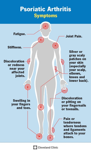
It’s also not uncommon for the underlying immune disorder that causes Psoriasis to also cause additional systemic disease in other organs such as joint arthritis, inflammatory bowel disease, cardiovascular disease and other metabolic diseases because the inflammation doesn’t always stay localized just to the skin, especially in more severe cases (20-30% of cases). Recently the term “Psoriatic Disease” has been used to describe these more complex cases, driven by similar immune dysfunction, but not being localized to just the skin. Of these, joint arthritis (inflammation) is the most common escalation of skin Psoriasis, leading to a condition known as Psoriatic Arthritis. Most people are familiar with the term Arthritis, which generically refers to inflammation or irritation of the joint tissue (any joint). Psoriatic Arthritis specifically is a chronic disease that links skin Psoriasis immune dysfunction to inflammation in the joint tissue, causing swelling, irritation, tissue damage and potentially bone loss in severe cases. This can be especially debilitating, occurring in the fingers and toes (Distal interphalangeal), in multiple joints all over the body (Symmetric or Asymmetric), in the spine (Spondylitis) or in the hands and feet (Mutilans). The joint inflammation occurs in two categories, non-specific activation (unregulated cellular infiltration and inflammation mediated by signaling molecules and Neutrophils) and tissue targeting auto-immune disease (Tcells, Bcells and Antibodies that directly target joint tissue). Both can cause similar symptoms and damage, but the cell types and molecules involved can differ, leading to differential treatment approaches. A growing body of evidence suggests there are numerous gene mutations that increase the likelihood of one developing Psoriasis, especially HLA genes (involved in recognizing self vs foreign molecules), IL-12/IL-23 immune pathway as well as mutations affecting IL-17 and IL-22 Tcells. These discoveries support the rationale for targeting these pathways in treatment to help mitigate disease severity in patients suffering with Psoriatic Disease. The difficulty arises in identify which of the many immune mechanisms is driving the dysfunction, how out of balance they are in each patient and how much are other associated immune components involved (such as Th1 Tcells, Bcells, Natural Killer cells and gamma-delta Tcells). This variability in disease severity AND involved cell types is what drives the differences in how a patient is treated and what patients are treated with.

Common symptoms of Psoriatic Arthritis. Cleveland Clinic.

Because Psoriatic Diseases often include a more systemic component than Eczema, many preferred treatments target the specific immune pathways mentioned above systemically. I’ll break down the treatments into four categories; generic skin supportive therapies, topical anti-inflammatories, systemic corticosteroids and Disease-modifying antirheumatic drugs (DMARDs) that aim to slow down the immune response. Measures that promote overall systemic and skin health can help mitigate less severe Psoriasis symptoms, such as a balanced diet, skin moisturizing, short term UV/sun exposure and regular cleaning of affected areas. For Psoriasis that is primarily skin associated with no major systemic involvement, topical anti-inflammatory medications (both steroids and non-steroidal) can provide temporary relief to the irritation and skin damage during flares, though they aren’t typically recommended for long-term use. In moderate to severe cases, systemic treatments can provide better long-term remission in many patients, the challenge lies in which treatment is best for an individual due to the variability of disease. Again, systemic corticosteroid use can help to rapidly reduce inflammation but also serves to broadly suppress total immune function and as such is not recommended for long-term use. Other Disease-modifying antirheumatic drugs (DMARDs) such as methotrexate and cyclosporin can help to reduce systemic over-activation of the immune system, but since they are broad acting and not immune pathway specific they can also lead to immunosuppression and a reduced ability to fight off other diseases in some patients. In recent years new classes of immune pathway specific therapeutics have been developed to target the parts of the immune system that are commonly dysregulated in Psoriatic Diseases including; TNFa signaling molecules, IL-12/23 pathway, IL-17 Tcell pathway, JAK/STAT/Tyk2 (broader immune activating molecules) and Bcell/Antibody production. If you watch TV you’ve probably seen commercials for Skyrizi/Tremfya (IL-23 blockers) or Otezla (PDE4 blocker) recently, these are all newly approved medications for Psoriasis and related diseases. These types of treatments are designed to specifically block and reduce activity in a specific immune pathway in an attempt to try and bring the immune system back into balance, rather than broadly suppressing the whole immune system. While specificity can be very beneficial, the two challenges are identifying WHAT the right pathway to target is and mitigating the side-effects of blocking that immune pathway and other treatment associated side-effects. Unfortunately, in many cases identifying the most effective treatment with the fewest side-effects can be a challenging process, wrought with trial and error, changes in medication and evolving disease symptoms. This becomes a very personalized process that needs to be addressed on a case-by-case basis.

Concluding Remarks
Many of us know people suffering from some form of skin disease, with Eczema and Psoriasis being the two most common. While they share some common symptoms and treatments at a more basic level, they are driven by a wide range of different cell and immune dysfunctions that can require much more specific attention as the disease severity worsens. Treatment can range from basic cleaning and skin hydration to short-term anti-inflammatory treatments to long-term immune modifying therapies. And while these different treatments can help reduce symptoms and mitigate life altering challenges, management often becomes a life-long process for many suffering from these diseases. For long-term immune modifying treatments, the process of optimizing treatment can be challenging and frustrating. Whenever one attempts to modify an immune response, the body naturally creates a feedback loop and response to the treatment, and while for many these feedback loops to the treatment are minimal, sometimes this can create new challenges or immune dysregulation that needs to be understood and addressed. This process is confounded by the fact that currently our healthcare system doesn’t tend to prioritize personalized immune function testing to help identify an individual’s personal immune dysfunction and immune signature, slowing down the process of finding the optimal treatment. In the next posting I’ll be tackling the topic of “Personalized Medicine and Immune Signatures”, where the technology stands to making this a reality, how it might help guide an individual’s treatment, help us understand our own body function, but also where the challenges lie in understanding and applying the information gained. While there can be a lot of power in gathering individual personalized data, a single test rarely tells the whole story and with our ever evolving understanding of the human body there are still many gaps in this knowledge.

This content is for educational and informational purposes only and is not a substitute for medical advice. It does not provide diagnosis, treatment recommendations, or guidance for any individual’s specific medical situation.

References:
• Armario-Hita JC et al. Updated Review on Treatment of Atopic Dermatitis. J Investig Allergol Clinn Immunology. 2023; 33(3): pg158-167.
• Brunner P et al. The Immunology of AD and its Reversibility with Broad Spectrum and Targeted Therapies. J Allergy Clin Immuno. April 2017; 139 (4 Suppl): pgS65-S76.
• Cleveland Clinic. Psoriatic Arthritis. https://my.clevelandclinic.org/health/diseases/13286-psoriatic-arthritis. 2025.
• Dall’Oglio F et al. An Overview of the Diagnosis and Management of Seborrheic Dermatitis. Clin, Cosmet and Invest Dermatology. Aug 2022; 15: pg1537-1548.
• El-Esawy FM et al. Methotrexate Mechanism of Action in Plaque Psoriasis: Something New in the Old View. J Clin Aesthet Dermatol. Aug 2022; 15(8): pg42-46.
• Emmungil H et al. Autoimmunity in Psoriatic Arthritis: Pathophysiological and Clinical Aspects. Turkish Journal of Med Science. Aug 2021; 51: pg1601-1614.
• Freitas E at al. Bimekizumab: The New Drug in the Biologics Armamentarium for Psoriasis. Drugs in Context. 2021; 10: 2021-4-1.
• Gossec L et al. EULAR Recommendations for the Management of Psoriatic Arthritis with Pharmacological Therapies: 2023 Update. Ann Rheum Disease. March 2024; 83: pg706-719.
• Kim J et al. Pathophysiology of Atopic Dermatitis: Clinical Implications. Allergy and Asthma Proceedings. March 2019; 40(2): pg84-92.
• Li M et al. Beyond the Dichotomy: Understanding the Overlap Between Atopic Dermatitis and Psoriasis. Front Immunology. Feb 2025; 16:1541776.
• Loiselle AR et al. Prevalence and Co-Occurrence of Eczema Types in Adults in the United States: Insights from the All of Us Research Program. J of Invest Dermatology. March 2025; 145: pg2360-2363.
• Lowes M et al. Immunology of Psoriasis. Annu Rev Immunol. 2014; 32: pg227-255.
• NIH National Cancer Institute SEERS Training Module. Anatomy of the Skin. https://training.seer.cancer.gov/melanoma/anatomy/. 2025.
• Orlando E, Nona L. Eczema Vs. Atopic Dermatitis: Causes, Diagnosis, Treatment. University of Central Florida. https://ucfhealth.com/our-services/dermatology/eczema-vs-atopic-dermatitis/#:~:text=Eczema%20and%20atopic%20dermatitis%20(AD),out%20of%20seven%20different%20types. 2025.

Intro to Immunology, 1/19/26

The Immune System is a complex interplay between a dozen+ cell types, signaling molecules, anti-microbial systems and physical barriers that serves to protect our body from internal and external invaders in addition to leading clean up and repair of natural damage that occurs as part of daily life. The goal of this writing isn’t to outline every detail and nuance of the Immune System, but rather to provide a general overview of the major pieces and parts that contribute to protecting our bodies, allowing us to navigate life in a complex world full of dangers. In addition to providing a few relatable real-world examples of how our immune system protects us, I will also include some information on how it can become dysfunctional in its own right, leading to self-inflicted harm. This article will be a precursor to many more detailed and disease specific articles focused on creating comprehensible and digestible understanding of complex disease topics that affect life.

TERMS:
Innate Immune System = Non-specific pieces of the immune system.
Adaptive Immune System = Pathogen specific immune system.
Mucosa = Soft tissue that lines organs such as the airways, intestines and reproductive tract, allowing regulated entry/absorption from the surroundings to the rest of the body.
Pathogen = Agents such as viruses, bacteria and parasites that can infect a person.
Antigen = Any material foreign (from the outside world) or naturally occurring in the body that can cause an immune response.
Anti-microbial = Something that is known to stop the growth of or kill microorganisms such as bacteria, viruses, fungi or parasites.

The Immune System is divided up into two separate arms, the Innate Immune System which is made up of quick reacting non-pathogen specific protection mechanisms and the Adaptive Immune System, comprised of antigen specific responses that can be both short lived and very long lived (memory). The Innate Immune System is made up of three arms; natural barriers to entry (such as skin and airways), circulating molecules that destroy invaders and damaged cells and immune cells that wander the body looking for things to clean up. All of these mechanisms function in a pathogen agnostic manner, meaning it doesn’t matter if the invader is Bacteria A or B, Virus A or B. Meanwhile the Adaptive Immune Response exists for when these initial mechanisms get overwhelmed and our body needs to create a more specific response in order to eliminate the threat before it can cause too much ancillary damage to our own body. T-cells (Helpers and Killers) and B-cells (producing Antibodies) are the two main components of the Adaptive System that get amplified in response to a specific target. Once the threat is cleared, a portion of these cells can remain dormant in our body for years, just waiting for the same target to return (our Immune Memory). If that’s all you are interested in learning about the specific cells and pieces of the Immune System, feel free to skip down to the sections on “Health, Immune Function and Dysfunction” below, as the next section is a more detailed and denser explanation of the Immune System’s parts and pieces (for the nerds out there).

Components of the Innate and Adaptive Immune System
Immune exposure and development begins in utero, but really kicks into full swing after birth, when we are thrust into a dirty world full of new antigens and pathogens. While the process of exposure and reaction to our environment never stops after birth, the human Immune System is mostly formed by 3-4years of age. The first line of defense of the Innate Immune System are natural barriers like the skin and mucosal cells lining the airways, intestines and reproductive tract. These create physical barriers that provide limited and restricted access to the body, helping to prevent bacteria, viruses and allergens from regularly gaining entry. In addition, hair in the nostrils along with mucus in the airways and intestines help trap outside materials, allowing the cells that line these surfaces to produce natural anti-microbial molecules that are capable of destroying some pathogens. If these external barriers and systems are damaged or overcome, then the body has two additional sets of quick responding Innate mechanisms that serve to clean up and protect our organs and tissues. The first of these are a series of circulating molecules that include anti-microbial peptides (similar to natural antibiotics), proteins designed to tag and identify foreign molecules (eg CRP, lectins and ficolins) and complexes that help destroy infected or damaged cells (complement proteins). None of these are living, but all are capable of interacting with their targets to assist in protecting our body.

Innate Immune Overview. Anaya JM et al. Autoimmunity, From Bench to Bedside.

The second set of Innate mechanisms comprise a diverse group of cells that are both programmed to both dispose of garbage and to signal other more specialized cells to enhance the immune response. All of these cells have Pattern Recognition Receptors that are designed to identify common patterns found on bacteria, viruses, parasites, environmental allergens and damaged cells. These common antigens are things that our bodies are exposed to daily, and that we’ve evolved to recognize and clear out as part of our normal immune function. Granulocytes are some of the first cells to respond to issues, these include cells such as; neutrophils that help eat up foreign invaders and destroy damaged cells, eosinophils that help kill parasites and mast cells and basophils that are commonly involved in responses to allergens. The next class of cells collectively known as phagocytes (Monocytes, Macrophages and Dendritic Cells) serve two important functions, to eat up and destroy any garbage they find floating around and in more extreme cases to use this garbage to activate more specific Adaptive Immune responses (remember this in the next section). The last subset of Innate Immune cells are specially programmed killers, whose sole goal is to destroy foreign invaders and mutated cells (ie cancer cells). They are responsible for maintaining the balance between normal growth and dysfunction (parasites, mutations and cancers), preventing the latter from coopting our body.

Primary cells of the Adaptive Immune System. https://cellcartoons.net/lymphocytes/

While the Innate Immune System has some generic patterns it recognizes, it is NOT responding in a pathogen specific way. Meaning it would respond to most bacteria using similar mechanisms and the same goes for many similar viruses. If the first lines of defense fail to contain the pathogen/damage, cells known as Antigen Presenting Cells (often Phagocytes) are able to trigger activation of the pathogen specific Adaptive Immune Response. The two cells many people are familiar with that drive Adaptive responses are T-cells and B-cells. There are three major classes of T-cells, each serving a different role in enhancing specific immune responses; CD4+ T-helper cells, CD8+ Cytotoxic T-cells (specialize in killing) and gamma-delta T-cells (Immune surveillance). During normal development, each CD4+ and CD8+ T-cell is created with a unique random target, in fact there are millions of these unique cells just floating around the body. If by chance one of the Antigen Presenting Cells finds a new infection that matches the unique target on one of these T-cells, then that unique cell multiplies in response to the specific activation signals. The CD4+ Helper T-cells role is to identify the location of these unique infections and to signal other killer cells and B-cells to migrate to the site of infection/damage, thus specifically amplifying the immune response. There are Helper T-cell types that target infections hiding inside our cells, others that target free floating infections, some that participate specifically in allergic responses, others that help in tissue repair and even some that are specifically programmed to slow down inflammation if things get too wild and crazy. Meanwhile the CD8+ Cytotoxic (Killer) T-cells have overlapping specificity to the Helper T-cells, but instead of recruiting other cells and amplifying immune response, their sole job is to kill any cell that matches their unique target. This includes both infected cells and mutated/cancerous cells.

The last arm of the Adaptive Immune Response is the infamous B-cell and the Antibodies they produce. Just like the T-cell, during normal times our body creates an army of millions of B-cells, each with a unique random target, just hanging around, waiting for a Phagocyte or T-Helper cell to show them the right target. If they find the unique antigen that matches their Antibody, they go into action, multiplying and cranking out Antibodies against the target. These Antibodies are basically very sticky keys (though shaped more like a Y), that bind to the infection/target, both marking it to be destroyed by the killer cells (discussed above) and walling off the target to prevent it from doing further damage to the body. In cases where the body amplifies T-cells and B-cells against a very specific infection/target, some of those cells mature and turn into memory cells that go into a hibernation like state, hanging out for years (or even our entire life). These cells lie in wait for that same infection/target to return, ready to quickly respond and prevent us from getting sick (this is generally how vaccines work). In total, our Immune System is a complex web or responses and interactions, all tightly regulated with feedback loops to allow careful responses to invading pathogens, repair damaged tissues and prevent excessive collateral damage to our own body from within.

Health, Immune Function and Dysfunction
You might be asking, how do all these cells and pathways affect how my daily bodily function, or when I get sick with a bacteria/virus, how about when things become dysfunctional leading to self-inflicted disease? Even more than the details above describing the different parts of the Immune System, these questions are much more pertinent to life on a day-to-day basis. Though unfortunately in some ways these questions are far more complicated than the Immune System itself. My goal in this section is to introduce some general trends and concepts, rather than to address every health condition, supplement, disease, Autoimmune condition out there. In subsequent posts I’ll be addressing specific diseases, conditions and health topics that are either of specific interest to me or that have been requested by people in my life (feel free to reach out with your request).

The Immune System is something that’s constantly working, reacting, resting and repairing the body every single day. The surrounding environment is full of agents that have the potential to harm us, cause disease or coopt our cellular machinery, and the Immune System is one of the major factors that keeps a lot of these in check. This includes preventing allergens from entering the body, killing off bacteria/viruses that we’re exposed to on a daily basis to repairing damaged tissue from all the cuts and scrapes we accumulate, the Immune System does a little bit of everything. Our external environment is constantly challenging our body’s defenses, and for the most part it does a good job of blocking out many things, killing off small numbers of invading pathogens, clearing out cancerous/dead cells and repairing both internal and external tissue damage. There are many aspects of our daily lives that help keep things functioning well, including proper nutrition, a good supply of energy (aka calories), adequate sleep, low stress and exercise. Even if we did everything perfectly to keep the body functioning at 100% (basically impossible), our body’s defenses and repair mechanisms sometimes need a little extra help.

Overview of the Infection cycle and Immune Response. Kuby Immunology.

The most common challenge our Immune System faces is an overwhelm by too many pathogens. If a pathogen is either too numerous or has mechanisms to evade the immune system, then our body starts going into overdrive, manifesting in what most of us think of as ‘getting sick’. Our body temperature increases (fever) to both inhibit pathogen replication and increase immune activity, our vessels flood immune cells to the site of infection (causing inflammation), we have aches and pains (caused by stimulation of local nerves and the battle that’s being waged around them) and we become fatigued as our body reallocates energy to fighting off the infection. This infection can happen locally around a cut/wound, in a specific tissue (like a stomach bug or respiratory infection) or systemically (in your blood) affecting the whole body. There are varied degrees of severity of this process, at it’s best our body responds quickly to control the invading pathogen and we barely feel any symptoms (vaccines help prime these responses to be quicker), but at it’s worst the invading pathogen spreads throughout our body both infecting and killing cells directly but also hyper-activating our immune system so much so that it causes self-inflicted damage in its effort to clear the infection (collateral damage). So in general it is good to let the body go through the inflammation and healing process naturally, though if a fever gets too high or the immune response too severe, it can start to damage organs and cause more harm than good, and that’s when it’s a good idea to seek medical attention (especially for younger children). Unfortunately, sometimes it’s hard to know when our body crosses this line and what the best line of care is, because how each of us responds and how much each individual can tolerate is different.

The other challenge our immune system regularly faces is trying to keep the body from dysfunctionIng and directly attacking itself. This can occur in two main forms, Autoimmune disease (where the body mistakenly attacks itself) and Cancer (where mutations cause uncontrolled growth). Autoimmune diseases cover a wide-range of issues including Rheumatoid Arthritis, Type 1 Diabetes, Eczema and Psoriasis, Crohn’s disease, Multiple Sclerosis and Lupus among many others. In all of these diseases, some part of the Immune System becomes misdirected and hyperresponsive, leading to the body attacking itself and damaging a specific tissue or cell type. These diseases can be triggered by genetic factors, infections that cause a misfiring of the Immune System, random mutations causing auto-reactive cells, environmental triggers, chemical damage and other factors that we’ve yet to identify. Because of the varied causes of Autoimmune Diseases, we can’t always predict who or when they might occur. Even more challenging is that not all Autoimmune Diseases of a single classification are created equal, meaning one person with Eczema (skin disease) might have a different pattern of immune dysfunction than another person, so what treatments and interventions work best of each could be different (coming in the next blog). These are the types of discussions I’ll be trying to dive deeper into as part of future writings, discussing the more subtle nuance of a single disease or classes of diseases and what that means for those working through those challenges.

The other major classification of dysfunction is the diverse field of Cancer Biology. In over-simplified terms, Cancer is caused by a mutation that occurs somewhere in the body (really could be anywhere) that creates cells that divide, grow and spread in an uncontrolled manner. These cells can be in Immune Cells, epithelial cells, structural cells, nerve cells and so on. Regardless of the cell type, during normal maintenance processes, the Immune System is responsible for identifying these malfunctioning cells and destroying them before they can divide and spread, but when they are missed during the normal surveillance process is when disease can happen. These mutations can lead to the growth of either benign or malignant tumors, the former being a non-cancerous clump of cells (don’t spread and invade other tissues) and the latter growing rapidly and spreading throughout the body causing serious risk to normal tissue function. While there are some patterns in the mutations/cells that cause Cancer, there is also a lot of variability, which adds to the challenge of treating the disease, especially in the context of minimizing damage to non-cancerous tissue in the body, not always an easy feat. As such there is a wide range of therapeutic options, ranging from targeted radiation that kills the cancerous cells (and nearby cells), drugs that can interrupt cell division and growth, specialized therapies that can tag cancer cells for the immune system to identify (if they are unique enough) and the use of specially designed synthetic immune cells specially designed to attack the cancerous cells (one of the newer technologies). While the Oncology field has made great strides in understanding and treating Cancer, there are still many cancers that are hard to predict, identify and treat, meaning much more research is needed to improve the understanding of the what, how and why.

The process of healthy and normal immune function is a complex web of maintenance, reaction and regulation. Serving to protect our bodies, maintain normal growth and fix damage. All of the information above is just a small window into the complexities of disease and immune functionality. It’s also important to keep in mind that we’re all individuals and while I’ve outlined some general patterns that apply to most humans, we each have our own unique challenges, strengths and body types that need to be treated as such. This blog series is intended to be an ever-evolving work that serves to help those with Non-Scientific backgrounds further understand complex health related topics. As such I welcome all questions and any constructive comments and critiques from Scientist and Non-Scientist alike. Up next, Skin Diseases, Disruption and Immune Dysregulation…. If you’re a more visual learner, check out the University of California Televisions video on Immunology Basics, it’s about 1.5h long, so strap in.

This content is for educational and informational purposes only and is not a substitute for medical advice. It does not provide diagnosis, treatment recommendations, or guidance for any individual’s specific medical situation.

References

  • Abbas A et al. Cellular and Molecular Immunology. Elsevier Science, 2003.
  • Alberts B et al. The Cell. Garland Science, Taylor and Francis Group. March 2002.
  • Alotiby A. Immunology of Stress: A Review Article. Journal of Clinical Medicine, 2024; 13, pg6394.
  • Anaya JM et al. Autoimmunity, From Bench to Bedside. El Rosario University Press. September 2013.
  • Goldsby R et al. Kuby Immunology. WH Freeman & Company. January 2002.
  • Jain N. The Early Life Education of the Immune System; Moms, Microbes and (missed) Opportunities. Gut Microbes, Sept 2020.
  • Shishido SN et al. Humoral Innate Immune Response and Disease. Clinical Immunology, June 2012; 144, pg142-158.
  • Wrotek S et al. Let Fever do it Job. Evolution, Medicine and Public Health, Nov 2021; pg26-35.

Catching Up with Covid, 11/12/20

Topics:
Vaccines
Therapeutics
Reinfection and Immunity
Children and Schools
Looking Forward


Well, a lot has happened since I last wrote a Covid related piece, and a lot of important things have come to light very recently. In the past six months those of us in the United States have endured several waves of outbreak, and currently as of this writing we’re in the middle of the worst outbreak since the initial outbreak in April/May (Nov 11th, 2020; 144,000 New Cases, 65,000 hospitalized patients and 1400 New Deaths, John’s Hopkins data). There are many possible reasons for this proliferation in infections, but it’s not specific to a political party, demographic or portion of the country. Rather than argue about that, I’ll simply say, it doesn’t seem like people are acting responsibly and we’re going to pay the price, as death rates always lag a few weeks behind infections.

In the following paragraphs I’ll outline a lot of what has happened with the development of the SARS-CoV-2 Vaccines (including immunity, data, side effects, and timelines), what we’ve learned about the virus and how we’re treating it with new therapeutics, what the data tells us about how safe schools are and lastly some additional important notes about transmission. Note that this write-up is based on the information known as of 11/11/20, and I’m sure in the weeks following a lot of new information will come out. So let’s dive right in to the topic on almost everybody’s mind….are the vaccines going to work, when will they be approved and how long before ‘I’ can be vaccinated?

SARS-CoV-2 Vaccine Development and Trials:

The big headline on 11/9/20 was that interim data on the Pfizer/BioNTech’s vaccine “BNT162b2” shows 90% efficacy in preventing SARS-CoV-2 infection. Before we jump to too many conclusions, let’s take a step back and talk about how we got where we are and what we know. Since the release of the first genetic sequences of SARS-CoV-2 way back in January 2020 (10 months ago!) the scientific community has been working at a feverish pace to learn everything we possibly can about the novel coronavirus, turning out a mountain of research that would normally take a decade, in just a year’s time. A search for scientific articles containing “Covid” on pubmed.ncbi.nlm.nih.gov/ returns 68,494 hits! On top of this, many of the biggest Biotech companies in the world have dedicated huge proportions of their of their staff (often working lots of overtime) to the single task of solving this public health crisis, so believe me when I say this has been a truly unprecedented time for Science.

Vaccines against SARS-CoV-2 have progressed along timelines never seen before, partially because of this massive effort by the scientific community to execute multiple steps in the pipeline simultaneously, building off previous research on SARS-CoV and utilizing advances in vaccine development discovered in recent years. But questions remain, will the vaccine work, what kind of immunity is possible against SARS-CoV-2 and how long will it last? A lot of research has focused on assessing two major pieces of the human memory immune response to viruses; Bcells/Antibodies and Tcells. Most people have heard of antibodies (and the Bcells that produce them), and while most of the literature has shown that moderate to severe cases of Covid-19 lead to strong neutralizing antibody responses (Long April 2020), the durability (how long they last) is still open for debate and seems to depend on the severity of the disease experienced (Long June 2020). Thankfully the other arm of the memory immune response (CD4+ and CD8+ Tcells) seems to be far more robust and durable, and many studies have shown strong levels of anti-viral activity in a wide array of Covid-19 patients, even those who become seronegative (negative for antibodies) (Sekine June 2020, Le Bert July 2020, Grifoni June 2020). So this brings us back to vaccines, and a growing mountain of evidence that a well designed vaccine that leads to a robust immune response can subsequently lead to immunity against Covid-19.

There are currently 10 vaccines in Phase 3 clinical trials (raps.org), this is the final Phase of testing in which a large number of patients are vaccinated with the aim of looking at how effective the test vaccine is at preventing SARS-CoV-2 infection compared to a placebo control. Each trial is set to vaccinate upwards of 60,000 of people and won’t close down until a certain number of people become infected (doesn’t matter if they’re placebo or test vaccine). The threshold for the Pfizer trial is set at 164 infections, and the trial currently sits at 94 positive Covid cases, meaning they’re targeting another 70 positive Covid infections in the study before they close down and analyze all the data (though this could happen sooner). Early data from Phase 1&2 Trials of the successful vaccine candidates have shown a strong and consistent induction of the immune system, with SARS-CoV-2 specific antibody titers and SARS-CoV-2 reactive CD4+ Tcell levels being as high or better than those found in patients who have recovered from natural Covid-19 disease (Folegatti July 2020, Anderson Sept 2020, Sahin Sept 2020, Walsh Oct 2020). While the long term efficacy (>1year) of these responses is not known at this time, these trials are set to follow patients out for 2 years post-vaccination, and so far early returns (3-4months post-vaccination) are all promising that the vaccines create durable responses that will last for at least some time (TBD).

So we might have an approved vaccine before the end of the year (maybe even several), what next? Well thankfully for the general public many of these companies have already started building production capacity and scaling up production, banking on a successful Phase 3 trial (a big gamble). This means that if/when their vaccine is approved they are ready to start shipping vials almost immediately. Both the Pfizer/BioNTech and Moderna vaccines are mRNA vaccines, a relatively new technology that is MUCH faster to produce than traditional protein based vaccines. Problem is, there are some 7.5billion people to vaccinate, and initial estimates from Pfizer are to have enough vaccine for 50million doses (25million people) this year, that’s not a lot when distributed around the world. The WHO has a plan to evenly distribute the vaccine across all countries, but it remains to be seen if wealthier nations allow this to happen (WHO.INT/). Here in the US a team led by the Army have already worked out a supply chain and distribution plan to the different states, and then it’s up to each state to create a prioritization plan on who gets vaccinated first. Here in Colorado a rough draft of this plan has already been submitted to the CDC for approval, check this link for the full draft.
Skip to page 22-23 if you want to see the tiered priority list of who will be vaccinated first. In short the higher risk professions and people will be given first priority, even then estimates are that Colorado will receive 100,000 doses in the first shipment, enough to vaccinate 50,000 people, only a portion of those in Tier 1A. For those of you in the healthy general public, expect to wait until next Spring/Summer to get vaccinated (these timelines are still very fuzzy).

So whenever your turn comes up, there is a big question of what to expect from this vaccine. First off, there might be several choices available, and it remains to be seen if they all have similar efficacy and longevity. Second, there are different kinds of vaccines (mRNA, whole inactivated, subunit vaccines, hybrid vectors, etc) (Krammer Sept 2020). We don’t yet know how they stack up against each other, so I won’t elaborate any more at this time on them, but welcome questions about the different types if anybody has them. None of these vaccines are using live replication competent virus, so they do NOT cause infection. Two things to note about the vaccines in full disclosure, it’s not going to be as convenient or as comfortable as other vaccines we’re given in a more routine manner. This is part of what was sacrificed to make these timelines possible. Note, this DOES NOT in any way mean safety corners were cut or that regulatory steps were skipped, but rather it means that the comfort and patient experience isn’t quite as clean or easy. Many patients receiving the two dose vaccine regimen (Oxford, Moderna, Pfizer) report mild to moderate flu like symptoms within 24hours of administration; fever, fatigue, headache, etc. The symptoms are reported to go away within 24hours and are indicative of the body mounting a very strong immune response against a very strong vaccine, a good thing! Just be prepared to take a day off after receiving the vaccine, because there’s a good chance it’ll knock you on your ass (very short term).

Therapeutics

Other big news that came out this week was that Eli Lilly’s monoclonal antibody therapy against Covid-19 was given Emergency Use Authorization, though it is only being prescribed to elderly and high-risk patients, but at no charge (per US Government). Thankfully this is one of several therapeutics that have been shown to be efficacious in reducing the severity of Covid-19. Therapeutics can be divided up into those that act during the early stages of infection aimed at reducing the viral load and those acting during the later stages of infection that aim to minimize the damage caused by the immune response. In the former are anti-viral drugs such as Remdesivir (Gilead) (Spinner Aug 2020), monoclonal antibodies like Bamlanivimab (Eli Lilly) and REGN-COV2 (Regeneron, still in Phase 3) and a host of other anti-virals and biotherapeutics progressing through clinical trials. Convalescent plasma, while initially promising and helpful for some patients, is becoming less commonly used due to the inconsistent levels of neutralizing antibodies in donor patients (Agarwal Oct 2020). All of these treatments have been shown to be most efficacious when administered during the early stages of infection, before the disease reaches critical levels. Once the infection becomes more severe, requiring hospitalization, most of the therapeutics above have been shown to have minimal impact on the cytokine storm induced pathology, but this is when dexamethasone comes into play. A corticosteroid first approve in 1958, it acts as an immunosuppressant to help control the body’s over-reactive immune response to the virus that causes much of the late stage disease pathology, keeping many patients off ventilators (Tomazini Sept 2020). While none of these therapies are cures they have been instrumental in helping save some patients and in reducing the overall mortality rate. THIS is what the lockdowns did, they bought the Medical and Scientific community time to catch-up so that we could save more lives, though we still have a long way to go.

Reinfection and Immunity (Update)

Another big question that has arisen recently is about the potential for long term immunity and reinfection, the latter was recently proven by several case studies in Nevada, Hong Kong, Belgium and Ecuador (Iwasaki Nov 2020). First let’s start with what’s been learned about the immune response (for a more details on the immunology/virology, see my previous post). SARS-CoV-2 is a fairly sinister virus, in addition to asymptomatic/pre-symptomatic spread, the immune responses caused by it are quite variable. In many asymptomatic and mild cases the levels of neutralizing antibodies and length of seropositivity were shown to wane quite rapidly (Long June 2020), while those who had more severe disease had longer lasting humoral responses. Thankfully as I briefly introduced above, Tcell immunity seems to be more consistent across all infected patients (Sekine June 2020) and plays a major role in the memory response to the virus. Research has also shown that some people who have been previously infected by other coronaviruses have cross reactive memory responses to SARS-CoV-2 (Sette Aug 2020, Mateus Aug 2020), and that children may harbor higher levels of these cross reactive antibodies, a potential reason they are more resistant to Covid-19 (Ng Nov 2020). Preliminary data from one paper also showed that immune priming using the seasonal influenza vaccine can help promote immune responses to SARS-CoV-2. This is very preliminary lab data, and IS NOT inducing specific responses, but rather just an immune priming effect, similar to an adjuvant (Debisarun Oct 2020). This cross reactivity from other coronaviruses and immune priming from the flu shot DO NOT confer immunity, but may be part of the reason some people have more mild disease than others, though these hypothesis are still under investigation.

But if the body creates all these immune responses (some durable), how are people getting reinfected? Unfortunately for the handful of confirmed case studies we (Scientist) don’t know the exact answer. It’s possible the individual immune response was incomplete and thus proper memory cells were not created or maybe they were infected with such a high dose the memory response was overwhelmed? Thankfully, while viral sequencing of the Nevada case showed two separate viruses during the first and second infection, the mutations in their genomes did not appear to affect the antigenic sites the body recognizes; in short, mutation does not seem to be the reason for reinfection. Now, before you spin yourself into a frenzy, we’re talking about less than a dozen confirmed cases worldwide. If reinfection were a major issue (right now) we’d be seeing thousands of cases, not a handful, and these outliers were inevitable. So for now there is no need to panic about this topic, though it remains to be seen how long (beyond 1 year) immunity lasts and how stable the virus will be long term. So far due to proof-reading enzymes in the virus, and lack of selective pressures the virus seems to be fairly stable (Romano May 2020). Research on both these topics is ongoing, and as we get further out from the initial outbreak more will come to light.

Children and Schools

Now on to something a little more contentious, what role do children and schools play in the spread of the virus and is it safe to open up schools? I’ve spoken with many parents and teachers about this, and have heard stories on both sides; inability to work when kids are home, forcing a 6yo to do 5h/day of zoom (horrible), trying to educate one’s kids while working from home, teachers being given inadequate PPE to setup safe environments, teachers being guilted into working because if they don’t they’re responsible for our kids failing and on and on. It’s a terrible situation all around, so rather than focus on the politics, I’ll try to focus more on the research about how infectious are children and whether or not schools cause outbreaks.

By now most people are aware that children (0-19) rarely get severe disease and their risk of dying is very low. But the role of those under the age of 19 in spreading the virus is not so simple. Younger children (<4 or <6 in some papers) do not seem to be very strong vectors for Covid-19, meaning they are less likely to be infected and to transmit the infection to others. Older children (6-13) on the other hand were far more likely to become infected and transmit the virus, while young adults (14-19) experienced similar disease progression to people in their 20s (Goldstein July 2020). Though one study out of Duke did find that children of all ages (0-19) had similar nasopharyngeal viral loads (Hurst Sept 2020), but did not relate this back to infectious spread. So for children the story is more complicated because there does seem to be age related variability in regards to symptoms, though they can still become infected and transmit the virus in many instances.

Now the big question, what do the case studies from schools that have reopened show about the potential for Covid-19 outbreaks in schools? Unfortunately the answer again isn’t completely clear, partially because schools are reopening with a variety of mitigation measures, different levels of community spread and with different plans for testing, isolating and tracking outbreaks. Case studies out of Germany and Australia concluded that transmission within schools was fairly low IF community spread of the virus remained low AND rapid testing and contact tracing protocols were followed (Ehrhardt Aug 2020, Macartney Aug 2020). In both of these case the schools were running at reduced capacity, and following rigorous cleaning protocols, physical distancing and mask policies in some instances. Another case study out of Luxembourg found that during a community outbreak, secondary infections were transmitted throughout schools, though no large super-spreader events occurred in the schools (due to tracking and tracing programs) (Mossong Oct 2020). Overall the literature seems to agree that school related outbreaks are far more common in secondary and University level facilities (Goldstein July 2020), and while outbreaks do happen in younger children they do not seem to spread as rapidly. Measures such as reduced class sizes, physical distancing, rigorous cleaning, hand washing, mask wearing and testing and contact tracing will help limit any potential outbreaks. Though all of this is also contingent upon the level of community spread, and no study has tested or recommended reopening schools during larger and uncontrolled community outbreaks (like what’s happening in the US right now). The State of Colorado tracks all our larger outbreaks that are associated with a single facility/school, and if you look at the data you’ll notice many schools listed, but also many other businesses, events and gatherings.

Final Notes and Looking Forward

While there is a ton of promising data about vaccines, therapeutics, mitigation measures that help us control the spread, we are far from done with the pandemic. With cases, hospitalizations and deaths spiking all across the United States things are promising to get worse before they get better. Winter will bring the confounding issues of people being stuck indoors more often, influenza returning to the Northern Hemisphere (get your flu shot!) and the holidays (which promise to see lots of people traveling). If you have to travel I’d highly recommend you read my earlier post about navigating airlines, and be aware of the risks you are taking (United has fully booked flights right now, every seat). While airlines have been shown to not be high risk by themselves (Freedman Sept 2020), several outbreaks, including a large one on an Irish airline (59 cases), should be stark reminders of what can happen when people let their guard down (Murphy Oct 2020). Pandemic responses are not just about each person protecting themselves, but about all of us protecting those around us as well. Boulder County just went back into ‘Safer at Home: Level Orange‘, heavily restricting gatherings (2 households/10 people max) and further restricting other events and businesses. While we in the Scientific/Medical community are Catching up with Covid, it appears Covid is catching up with the general public….it’ll be a race to see who comes out on top.

Citations:
Agarwal et al, Convalescent plasma in the management of moderate covid-19 in adults in India: open label phase II multicenter randomized controlled trial (PLACID Trial). BMJ, Oct 22, 2020.
Anderson et al, Safety and Immunogenicity of SARS-CoV-2 mRNA-1273 Vaccine in Older Adults. NEJM, September 29, 2020.
Eli Lilly, Lilly’s neutralizing antibody bamlanivimab (LY-CoV555) receives FDA emergency use. Press Release, 11/11/20.
Debisarun et al, The effect of influenza vaccination on trained immunity: impact on COVID-19. medRxiv pre-print. October 16, 2020.
Ehrhardt et al, Transmission of SARS-CoV-2 in children aged 0 to 19 years in childcare facilities and schools after their reopening in May 2020, Germany. Eurosurveillance, September 10, 2020.
Folegatti et al, Safety and immunogenicity o ChAdOx1 nCoV-19 vaccine against SARS-CoV-2: a preliminary report of a phase 1/2, single blind, randomized controlled trial. Lancet, Vol 396: 467-478. July 20, 2020.
Freedman et al, In-Flight transmission of SARS-CoV-2: a review of the attack rates and available data on the efficacy of face masks. Journal of Travel Medicine, 1-7. September 18, 2020.
Goldstein et al, On the effect of age on the transmission of SARS-CoV-2 in households, schools and the community. medRxiv pre-print, July 26, 2020.
Grifoni et al, Targets of T Cell Responses to SARS-CoV-2 Coronavirus in Humans with COVID-19 Disease and Unexposed Individuals. Cell, 181; 1489-1501. June 25, 2020.
Guo et al, Long-Term Persistance of IgG Antibodies in SARS-CoV Infected Healthcare Workers. MedRxiv preprint, Feb 2020.
¬Han et al, Clinical Characteristics and Viral RNA Detection in Children with Coronavirus Disease 2019 in the Republic of Korea. JAMA Pediatric, August 28, 2020.
Hurst et al, SARS-CoV-2 Infections Among Children in the Biospecimen from Respiratory Virus-Exposed Kids. medRxiv pre-print, September 1, 2020.
Iwasaki et al, What Reinfections mean for COVID-19. Lancet Infectious Diseases, November 6, 2020.
Kaur et al, COVID-19 Vaccine: A comprehensive status report. Virus Research, Vol 288. August 13, 2020.
Krammer et al, SARS-CoV-2 vaccines in development. Nature, September 23, 2020.
Le Bert et al, SARS-CoV-2 specific T cell immunity in cases of COVID-19 and SARS, and uninfected controls. Nature, Vol 584, August 20, 2020.
Long et al, Clinical and immunological assessment of asymptomatic SARS-CoV-2 infections. Nature Medicine. June, 18 2020.
Macartney et al, Transmission of SARS-CoV-2 in Australian educational settings: a prospective cohort study. Lancet Child Adolescent Health, August 3, 2020.
Mateus et al, Selective and cross-reactive SARS-CoV-2 T cell epitopes in unexposed humans. Science, August 4, 2020.
Mossong et al, SARS-CoV-2 transmission in educational settings an early summer epidemic wave in Luxembourg. Pre-print, October 2020.
Murphy et al, A Large National Outbreak of COVID-19 Linked to air travel, Ireland, Summer 2020. Eurosurveillance, Oct 6, 2020.
Ng et al, Preexisting and de novo humoral immunity to SARS-CoV-2 in humans. Science, November 6, 2020.
Romano et al, A Structural View of SARS-CoV-2 RNA Replication Machinery: RNA Synthesis, Proofreading and Final Capping. Cells, Vol 9(5): 1267. May 20, 2020.
Sahin et al, COVID-19 vaccine BN162b1 elicits human antibody and Th1 T cell responses. Nature, Vol 586. October 22, 2020.
Sekine et al, Robust T cell immunity in convalescent individuals with asymptomatic or mild Covid-19. bioRxi preprint. June 29, 2020.
Sette et al, Pre-existing immunity to SARS-CoV-2: the knows and unknowns. Nature Reviews, Vol 20: 457-458. August 2020.
Spinner et al, Effects of Remdesivir vs Standard Care on Clinical Status at 11 Days in Patients with Moderate COVID-19. JAMA, August 21, 2020.
Tomazini et al, Effect of Dexamethasone on Days Alive and Ventilator-Free in Patients with Moderate or Severe SARS Distress Syndrome and COVID-19. JAMA, September 2, 2020.
Walsh et al, Safety and Immunogenicity of Two RNA-Based COVID-19 vaccine candidates. NEJM, October 14, 2020.
Willyard, Ageing and COVID Vaccines. Nature, October 15, 2020.

COVID-19; Immunology and the Infection Cycle, 4/22/20

Sorry for the delay, but this one has taken a lot of time and thought to put together (and reading some 80 odd Covid research papers). My goal in this edition of Covid Science Theater 2020 is to talk about what happens when the virus enters our body, infects our cells and subsequently leads to either mild disease or more severe infections. It’s going to be a fairly dense article, but I’ll do my best to keep the science and terminology to something generally understandable and hopefully educational.

For those who don’t want to delve too much into the specifics of all the virology, immunology and pathology I’ll provide a short 1 paragraph set of clifs notes here. The virus most commonly enters the host through mucus membranes (eyes, nose, mouth) and infects vascular endothelial cells and cells of the lungs, kidneys, GI tract and begins to replicate. The body initially responds to the virus through a host of Innate Immune mechanisms; these generic counter measures are deployed against all invading pathogens as a first line defense and are not specific to the invading pathogen. Unfortunately, these initial immune responses aren’t always adequate to contain the virus (the virus sometimes evades destruction, other times the virus just overpowers the immune response) so our body deploys a second type of response known as the Adaptive Immune response. In this phase, T-cells and B-cells are primed to respond to the specific infectious agent (here, SARS-CoV-2). Often this two-pronged approach works to contain the infection, eliminate the virus and build up lasting memory to subsequent infections. Unfortunately in some people the virus spreads too rapidly and the immune response doesn’t respond appropriately, leading to destruction of their organs (notably the lungs) and potentially death. Sometimes this more severe outcome is caused by the virus itself, but more often it seems to be caused by an overzealous immune system trying to play catch-up. So there’s the quick and dirty; in the following paragraphs I’ll go into more detail about Viral Entry/Binding/Replication, Early Cellular Responses, Clinical Symptoms, Adaptive Immune Response and What Happens and Why the Immune System Sometimes Fails.

The information in the following paragraphs comes from a combination of basic immunology principles (Kuby Immunology textbook), observations and early research released about Covid-19 and conclusions drawn from earlier studies of SARS-CoV-1 (a virus that is very similar to the current SARS-CoV-2, but with some caveats of course). As Covid-19 is still a new disease, we are constantly learning new things about the virus, infection cycle and pathology, so while what I outline here is based on a lot of research, there are definitely aspects of this virus that we don’t fully understand, and need further investigation.

Graphic of the structure of a SARS virus, the S, M and E proteins are the most important in regards to host recognition, Li et al 2020.

Viral Entry and Replication

Coronaviruses get their name from the hallmark shape, a circular capsid (or shell) that is spiked with proteins on the outside and sheltering the virus genetic sequence on the inside. The Spike (S), Membrane (M) and Envelope (E) proteins make up the majority of the viruses outer shell, while the Nucleocapsid (N) protein found inside the virus assists in viral replication. This small assortment of proteins, plus a few others, make up the bulk of the very simple viral structure (Weiss 2005, Li 2020). The novel coronavirus 2019 (COVID-19) shares a lot of homology or similarity with the original SARS virus that was discovered in 2002, genetically 80% similar, while being 76-95% similar for the major proteins listed above (Xu 2020). This allows researchers to draw a lot of conclusions from previous research on SARS-CoV-1, though we must be careful when doing so, as there are some known (and unknown) differences between the two viruses. The infection cycle starts with the virus gaining entry to the host, usually through mucus membranes of the eyes, nose and mouth. Once inside the virus often begins it’s infectious cycle by infecting vascular endothelial cells that line vessels throughout the body. While different viruses have different mechanisms by which they enter host cells, SARS-CoV-2 binds to the ACE2 receptor using its spike protein (same as SARS-CoV-1), allowing it to enter the host cell (Jia 2005, Walls 2020). Like most viruses, SARS-CoV-2 then goes through a multi-stage process by which it hijacks some of machinery inside our own cells to in order to replicate, escape and subsequently infect more cells in a continual cycle (Frieman 2008).

Overview of the SARS viral life cycle inside the host, Frieman et al 2008.

Early Cellular Response

Thankfully our body has a whole host of immune mechanisms it utilizes to deal with infectious agents of all types. Almost as soon as an invading pathogen has infected our cells the immune system starts going to work. The Innate Immune response is our constantly active sentinel, whose cells are constantly circulating all over our body just looking for foreign invaders to attack and kill. These innate cells use Pathogen Associated Molecular Patterns (PAMPs), or markers of foreign invaders, as the initial signals something is wrong and that it’s time to go to work (Li 2020). Some cells go to work directly attacking the virus and infected cells in an attempt to destroy the virus, others release signaling molecules known as cytokines and chemokines that recruit other cells to help in the fight (Frieman 2008), and some cells just go ahead and sacrifice themselves in an effort to prevent the virus from hijacking them, a process known as apoptosis (Lim 2016).

Symptoms: What and How They Manifest

While this system works well for many invading pathogens (why we are not sick all the time), allowing our body to control the infection, many viruses (and bacteria) have evolved mechanisms by which to evade, subvert and co-opt the immune response to their advantage. For SARS-CoV-2 it seems to be able to prevent the host immune system from activating one of it’s key anti-viral signaling pathways, the Type 1 Interferon pathway (Lim 2016, Li 2020, Frieman 2008). While it is not known exactly how the virus subverts this system a few hypotheses involve the Nucleocapsid protein (Lim 2016), other non-structural proteins (Lim 2016), and some of the SARS enzymes (Chen 2014). So by reducing the host immune response the virus is able to more effectively replicate and spread, leading to a more systemic infection. This is when we start to experience more of the hallmark symptoms of the infection; fever, sore throat, coughing, fatigue, pulmonary inflammation leading to shortness of breath and possible pneumonia and lymphopenia (a decrease in lymphocytes, more on that later) (Huang 2020, Zhu 2020). Most of these symptoms are a physical outcome of the body’s ongoing fight with the virus, trying to delicately balance destroying the invader, while preserving the host organs and system. The fever is the immune system’s attempt to raise the core temperature enough to burn out the infection. The sore throat/cough is an outcome of our immune system attacking infected cells of the airways and trying to expel the invader (mmm mucus), same for the pulmonary issues (initially, more on this later too). While many of these symptoms may be scary and uncomfortable they are often a normal part of our body’s healing process when dealing with a foreign invader. So under normal circumstances, it’s best to rest and let your body do it’s thing, unfortunately this doesn’t always go as planned, as we’ll find out in the following sections….

Adaptive Immune Response; Stage 2

In the previous two sections you’ve seen how our well intentioned Innate Immune system can sometimes fail leading to illness, thankfully the body has a backup, the Adaptive Immune response. This secondary wave of the immune response goes into action very soon after the initial infection (several hours to few days, infection dependent) and is mostly comprised of two cell types; T-cells and B-cells. When the levels of virus in the body start to rise, several of the innate immune cells can act as activators of the adaptive immune response, taking pieces of the virus to specialized activation centers know as lymphoid organs. These centers of immune activation are spread all over our body and are the primary site of pathogen specific antigen (virus pieces) presentation. The antigen presenting cells (Dendritic cells are most prominent) present the virus to the T-cells and B-cells as if locks in a door, allowing the T-cells and B-cells to go to work making specific keys (receptors and antibodies) that can attack and destroy the pathogen in a very focused manner. The outer proteins that make up the viral capid (proteins S, M, E) tend to be the most effective as this is what is visible to our body when intact virion are released (Liu 2017). So the body makes a whole army of these specific cells that traffic to the sites of infection; T-cells directly attack the virus and infected cells, while B-cells make antibodies that bind to parts of the virus, preventing them from entering new cells and marking them for destruction (Liu 2017).

These two arms of the Adaptive Immune response are also what comprise our immunological memory. Virus specific T-cells and antibody producing B-cells remain dormant in specialized lymphoid organs (sometimes they also remain in circulation), just waiting for the virus to turn up a second time. This time since they are already primed and ready to go, memory T-cells and B-cells start attacking the virus almost immediately, usually preventing the virus from spreading and preventing us from getting sick. Studies of SARS-CoV-1 have found both memory T-cells and memory B-cells (producing neutralizing antibodies) that are capable of rapidly responding to viral reinfection (Li 2020, Liu 2017, Channappanavar 2014). In human patients who recovered from SARS-CoV-1 infection anti-SARS antibodies and memory T-cells were found in most patients up to 24 months after infection (Liu 2006, Ka fai 2008, Liu 2017). While antibody responses did decline over time in SARS-CoV-1 patients (many undetectable at 6 years), memory T-cell responses were conserved for up to 11 years after infection (Tang 2011, Ng 2016, Liu 2017). Similar high quality neutralizing antibodies have been found in COVID-19 patients, but since the disease is so new the longevity of memory responses to this new virus aren’t exactly known. Encouragingly, since SARS-CoV-2 is so similar to the original SARS virus, and lab testing has even shown that their be might cross-reactive protection between the two diseases (Walls 2020), there is much hope that the long lasting memory responses seen for SARS-CoV-1 would also apply to those who have recovered from COVID-19. All of this evidence, both old and new, does inspire a lot of hope that a functional vaccine would both be likely and very effective in providing some duration of immunity from COVID-19, but how long remains to be seen.

Graphical overview of the many cells and pathways involved in the host immune response to SARS. It’s a complex set of feedback loops and interactions with a lot of variables. Li et al 2020.

When the Immune System Fails, Severe Disease

The reason COVID-19 is such a scary disease, isn’t because our immune system has no problem fighting it off, but because in some percentage of the cases (uncertain, but estimates are as high as 10-20%) patients need to be hospitalized due to severe complications. If our immune system is so complex and so strong, why do patients with COVID-19 get so sick that they need hospital care? It comes down to numerous very subtle things this virus does that are different than coronaviruses that cause the common cold. One is the effect SARS-CoV-2 has on Type 1 Interferons mentioned earlier, reducing the body’s initial response to infection. Another early symptom seen in many severe cases is lymphopenia, or a loss of lymphocytes (notably T-cells) early on in disease (Huang 2020, Schmidt 2005, Weiss 2005). While the exact cause of this loss of T-cells is not known, it is hypothesized that the viral proteins may lead directly to T-cell death as a mechanism of immune evasion (Lim 2016, Li 2020). These mechanisms of avoiding immune detection along with the efficiency of viral replication can lead to an out of control infection very quickly.

But in the end, it’s only partially about the virus, and largely about an overexuberate immune response. In an attempt to catch-up to the wide-spread infection the immune response goes into overdrive, ramping up a lot of the inflammatory cells and signaling molecules that tell the body to attack the infection (Li 2020). This response does in fact kill the infected cells, but it also destroys lung tissue (primary target), vascular tissue, liver tissue and other infected tissues (Tian 2020, Schmidt 2005). This is often when the more obvious signs of pneumonia set in; the lungs fill with fluid, the efficiency of aveoli decreases (oxygen absorption) and breathing becomes very labored and difficult. This is the tricky thing about COVID-19, making our immune response more efficient would help prevent early infection, but later on would lead to increased tissue damage. But if we reduce the immune function of the body to prevent self-inflicted tissue destruction, we run the risk of allowing the virus to run rampant throughout our body. COVID-19 is a tricky disease to treat for these reasons, and because the disease severity has a wide range of outcomes for different people. In some, infection is very mild and asymptomatic, in others, their entire body shuts down as the virus (and immune system) destroys the host from the inside. The reason many comorbidities are important as risk factors for severe disease is that most of them either affect the immune system or lung function. Obesity, diabetes, auto-immune diseases all alter the immune system’s ability to function, making it harder to fight off the virus. COPD and asthma (though less prominent then thought) make the host pulmonary system more sensitive to damage caused by the virus and immune system.

But not all hope is lost! Because of the large body of evidence suggesting that SARS viruses create robust lasting immunity, this means a vaccine might be very effective at protecting most of the population. Also, now that there are many patients who have recovered from COVID-19, tests are underway to examine if using their plasma (containing antibodies) can help patients who are suffering from more severe cases of the disease (works for other viruses like Ebola). We also have several promising anti-viral agents that are already in clinical trials being tested against COVID-19, with hopes that one or more of them will help improve patient outcomes and be ready for use later this year. Unfortunately all of this does take time, meaning we won’t have a cure next month, but by slowing the spread of the virus, not only do we allow hospitals to manage the patient load, but we allow all the scientist out there to catch-up and produce much needed data, therapies and vaccines.

Thanks for reading. If you see any mistakes please bring them to my attention and I will correct them ASAP. If you have additional questions or want to discuss the immune response in more detail (this is a very high level overview) I’d be happy to do so via text or email. Stay safe and stay healthy.

Literature Citations:
Chan et al, Serological Responses in Patients with Severe Acute Respiratory Syndrome Coronavirus Infection and Cross Reactivity with Human Coronaviruses 229E, OC43, NL63. Nov 2005, Clinical and Diagnostic Laboratory Immunology.
Channappanavar et al, T cell-mediated immune response to respiratory coronaviruses. May 2014, Immunology Res.
Chen et al, SARS coronavirus papain-like protease inhibits the type 1 interferon signalling pathway through interaction with the STING-TRAF-3 TBK1 complex. Jan 2014, Protein Cell.
Frieman et al, SARS Coronavirus and innate immunity. 2008, Virus Research.
Huang et al, Clinical features of patients infected with 2019 novel coronavirus in Wuhan, China. Jan 2020, Lancet.
Jia et al, ACE2 Receptor Expression and Severe Acute Respiratory Syndrome Coronavirus Infection Depend on Different Human Airway Epithelia. Dec 2005, Journal of Virology.
Ka-fai Li et al. T cell responses to Whole SARS Coronavirus in Humans. Oct 2008, Journal Immunology.
Li et al. Coronavirus infections and immune responses. Jan 2020, Journal of Medical Virology.
Lim et al. Human Coronaviruses: A Review of Virus-Host Interactions. 2016, Diseases.
Lu et al. Immune responses against severe acute respiratory syndrome coronavirus induced by virus-like particles in mice. June 2007, Immunology.
Ng et al, Memory T cell responses targeting the SARS Coronavirus persist up to 11 years post-infection. March 2016, Vaccine.
Schmidt et al. Coronaviruses with a special emphasis on First Insights Concerning SARS. 2005, Birkhauser Advances in Infectious Diseases.
Tang et al, Lack of Peripheral Memory B cell responses in Recovered Patients with Severe Acute Respiratory Syndrome: A Six-year Follow-up Study. May 2011, Journal of Immunology.
Tian et al, Pulmonary Pathology of Early-Phase 2019 Novel Coronavirus (COVID-19) Pneumonia in Two Patients with Lung Cancer. Feb 2020, Journal of Thoracic Oncology.
Walls et al, Structure, Function and Antigenicity of SARS-CoV-2 Spike Glycoprotein. Apr 2020, Cell.
Weiss et al, Coronavirus Pathogensis and the Emerging Pathogen Severe Acute Respiratory Syndrome Coronavirus. Dec 2005, Microbiology and Molecular Biology Reviews.
Xu et al, Systematic Comparison of Two Animal-to-Human Transmitted Human Coronaviruses: SARS-CoV-2 and SARS-CoV. Feb 2020, Viruses.
Zhou et al, Clinical course and risk factors for mortality of adult inpatients with COVID-19 in Wuhan, China: a retrospective cohort study. March 2020, Lancet.